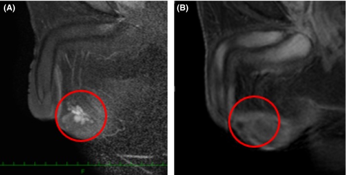
Figure 6.

A, Before glucocorticoid therapy, the testicular tumors demonstrated uniform enhancement, with discrete margins that were readily visualized on the T1‐weighted images following i.v. gadolinium administration. B, Three years later, the testicular tumors became undetectable on the enhanced T1‐weighted images
