
Fig 8. TAPF induces cFLIP and the levels of cFLIP increase concomitantly with protection against TNF + CHX.
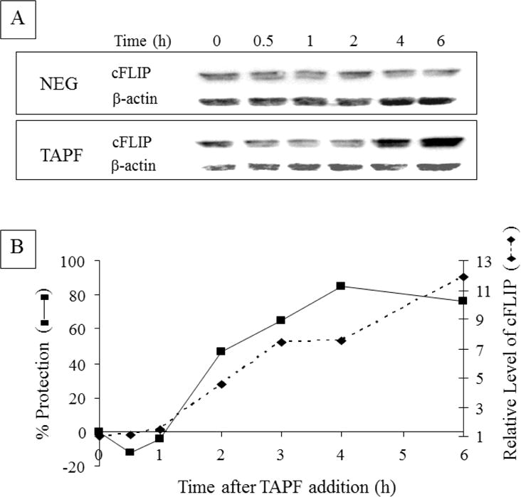
A: NHDF were either not treated (0h), incubated with TAPF or incubated with the negative fraction (NEG) for the times indicated. Cells were washed, lysed and equal volumes were analyzed by immunoblotting for cFLIP. B: Another experiment identical to A was performed. In addition, following identical TAPF treatment of samples in parallel for the times indicated on the abscissa, TNF+CHX were added and viable cells were stained 24h later. Percent protection was calculated as described for Fig. 1A. For each sample, n=3. Relative levels of cFLIP were calculated by first normalizing cFLIP to the corresponding β-actin signal; the time 0 value was set at 1 and values for all other times were reported relative to it. Data from one of three independent experiments are shown.