
Fig. 5.
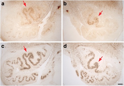
Degenerative changes in the patient’s left (a, c) and right (b, d) ION at a more rostral level, with immunostains to calbindinD28k (a, b) and GAD (c, d). A distinct region with more numerous calbindinD28k-labeled neurons is again seen in the dorsal arm of the principal olive bilaterally along with preserved GAD staining (red arrows). There is patchy loss of GAD staining, more prominent in lateral and ventral regions of the principal olive in this region of ION. Scale bar: 250 μm, A-D