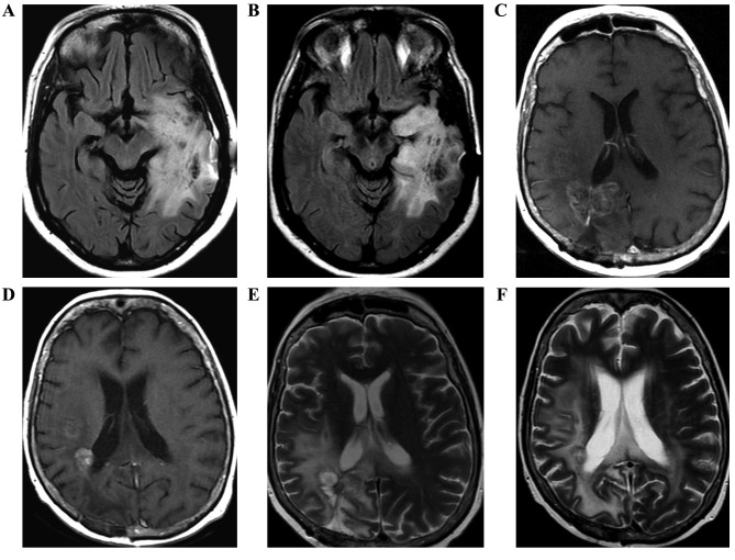
Figure 1.
Brain magnetic resonance images of three patients, with images prior to (A,C,E) and following (B,D,F) the commencement of treatment. (A) Axial flair shows part of the expansive lesion and the extensive perilesional edema in the left temporoparietal region, which extends along the left cerebral peduncle. (B) Axial flair in the control shows reduction of lesion dimension. (C) Axial T1W contrast-enhanced image demonstrates lesion with peripheral and irregular enhancement and perilesional edema. (D) Axial T1W contrast-enhanced image of the control with reduction of tumor volume following therapy. Postoperative changes in correspondence. (E) Axial flair demonstrates a heterogeneous lesion with hyperintense area. (F) Axial flair of the control shows reduction in the dimensions of the heterogeneous lesion with extensive perilesional edema. Magnetic susceptibility artefact by previous biopsy.

