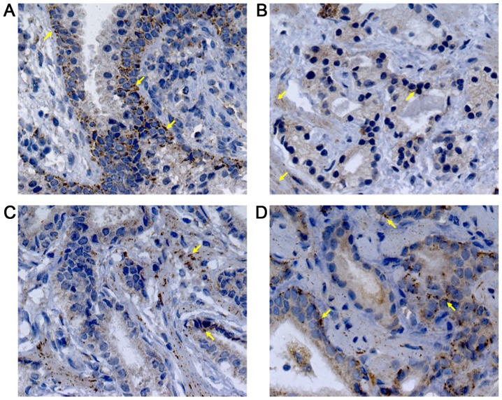
Figure 3.
Immunohistochemistry for PEDF in (A) BPH; (B) AdGS6, (C) AdGS7, and (D) AdGS8 biopsies. PEDF cytoplasmic expression in acinar and peri-acinar stromal areas was observed. PEDF staining was localized at basal cells (yellow arrows). Nuclei were counterstained with hematoxylin; magnification, ×400. PEDF, pigment epithelium-derived factor; BPH, benign prostatic hyperplasia; AdGSC, adenocarcinoma with Gleason score.

