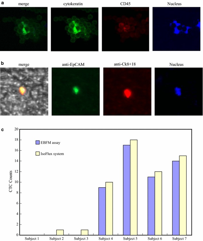
Fig. 8.

a Fluorescence images on the platform of the IsoFlux System, resulting from cytokeratin (green fluorescence), leukocyte (red fluorescence), and nucleus marker staining, and phase contrast image superimposed on the fluorescence image for an example of CTC recognition. b Fluorescence images on the EBFM assay, resulted from the FITC-conjugated anti-EpCAM (green fluorescence), Alexa594-conjugated anti-Ck8+18 (red fluorescence), and leukocyte marker (CD45) staining, and phase contrast image superimposed on the fluorescence image for an example of CTC recognition. c CTC counts for seven subjects, obtained using IsoFlux system, N50/P50 fibrous mats with anti-EpCAM grafts
