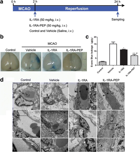
Fig. 1.

Effects of IL-1RA and IL-1RA-PEP on transient MCAO-induced Evans blue extravasation and BBB ultrastructure. a Experimental protocol. b, c Extravasation of Evans blue dye in brain tissues of the control, vehicle, IL-1RA, and IL-1RA-PEP groups was evaluated quantitatively. Data are shown as mean ± SEM; *P < 0.05, **P < 0.01, ***P < 0.001 compared to the vehicle group, #P < 0.05 compared to the IL-1RA group, n = 6 per group, based on one-way ANOVA with Bonferroni correction. d Ultrastructure changes in the BBB. The basement membranes (marked between black arrows in top panels) were observed in different groups. The black frames in middle panels are areas selected for amplification in the following bottom panels, to highlight tight junctions (pointed by white arrows). Scale bar, 500 nm and 2 μm for the inserts. MCAO middle cerebral artery occlusion, BBB blood-brain barrier
