Fig. 2.

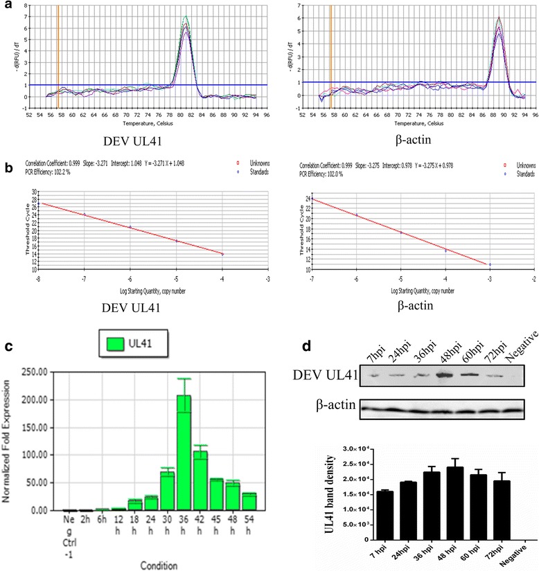
The transcription and expression kinetics of the DEV UL41 gene. a Melting curves of DEV UL41 (81 °C) and β-actin (89 °C). b Standard curves of DEV UL41 (Y = − 3.271X + l.048) and β-actin (Y = − 3.275X + 0.978), and UL41 gene (102.2%) and β-actin (102.0%) amplification efficiencies were approximately identical, with correlation coefficients of 0.999. c UL41 mRNA levels at 0, 2, 6, 12, 24, 36, 42, 45, 48, and 54 hpi were assessed by RT-qPCR and normalized toβ-actin. The UL41 transcript was detected at 6 hpi, increased gradually (6–30 hpi), reached its peak at 36 hpi, and then decreased steadily thereafter (42–54 hpi). d Representative western blotting results for UL41 at 7, 24, 36, 48, 60, and 72 hpi (up) (approximately 56 kDa) and quantification in the six groups (down). The negative group comprised mock-treated DEF cells detected with mouse anti-UL41 antibody serum (1:800) and goat anti-mouse IgG HRP-conjugated antibody (1:5000). The band density increased gradually (7-36hpi), peak expression occurred at 48 hpi, and expression decreased steadily thereafter (60–72 hpi). β-actin antibody was used to assess β-actin as the loading control
