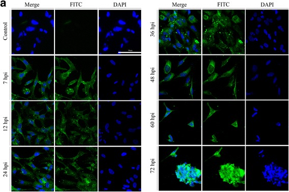
Fig. 4.

pUL41 intracellular localization in DEV-infected cells at different times. a Representative confocal immunofluorescence microscopic images of the UL41 localization (green in all images) at 7, 12, 24, 36, 48, 60, and 72 hpi. Nuclei are indicated in blue (DAPI). The control group comprised untreated DEF cells (magnification: 200×; scale bar: 50 μm) detected with mouse anti-UL41 antibody serum (1:800) and goat anti-mouse IgG (H + L) cross-adsorbed secondary antibody, Alexa Fluor 488 (1:1000). Significantly higher UL41 levels in the cytoplasm were evident in the DEF cells infected with wild-type DEV
