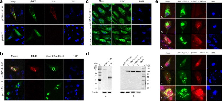
Fig. 5.

DEV-UL47 protein may translocate pUL41 into the nucleus in the absence of other viral proteins. All transfected samples were collected after 24 h post transfection. a DEF cells were transfected with pEGFP-C2-UL41 and pEGFP-C2, respectively. The EGFP-UL41 fusion protein was localized to the cytoplasm (green), further detected with mouse anti-UL41 protein polyclonal antibody (1:800) and goat anti-mouse IgG (H + L) cross-adsorbed secondary antibody, Alexa Fluor 568 (1:1000) (red). The pEGFP-C2 plasmid control was distributed throughout the cells (green). b DEF cells were co-transfected with pEGFP-C2-UL41 and pcDNA3.1-UL47. Much of the EGFP-UL41 fusion protein was localized to the nucleus (green), and pUL47 was mainly localized to the nucleus (red), detected with the rabbit anti-UL47 protein polyclonal antibody (1:1000) and goat anti-rabbit IgG (H + L) cross-adsorbed secondary antibody, Alexa Fluor 568 (1:1000). DEF cells were co-transfected with pEGFP-C2-UL41 and pcDNA3.1(+) as the control, and the EGFP-UL41 fusion protein was only distributed in the cytoplasm (green). c DEF cells were transfected with pcDNA3.1-UL47Δ40–50 and pcDNA3.1-UL47Δ768–777, respectively. A portion of the ΔpUL47 was localized to the cytoplasm (green), detected with rabbit anti-UL47 protein polyclonal antibody (1:1000) and goat anti-rabbit IgG (H + L) cross-adsorbed secondary antibody, Alexa Fluor 488 (1:1000). d Representative western blotting results for DEF cells transfected with pEGFP-C2-UL41 (approximately 83 kDa) and pEGFP-C2 (approximately 27 kDa), detected with rabbit anti-EGFP antibody (1:2000) (Beyotime, Shanghai, China) and goat anti-rabbit IgG HRP-conjugated antibody (1:5000). Western blotting results for DEF cells transfected with pcDNA3.1(+), pcDNA3.1-UL47Δ768–777 (approximately 87 kDa), pcDNA3.1-UL47Δ40–50 (approximately 87 kDa) and pcDNA3.1-UL47 (approximately 89 kDa), respectively, detected with rabbit anti-UL47 antibody (1:1000) and goat anti-rabbit IgG HRP-conjugated antibody (1:5000). The β-actin antibody assessed β-actin as the loading control. M. Precision Plus Protein™ Dual Color Standards. e DEF cells were co-transfected with pEGFP-C2-UL41 and pcDNA3.1-UL47Δ40–50, pEGFP-C2-UL41 and pcDNA3.1-UL47Δ768–777, respectively. Much of the EGFP-UL41 fusion protein was localized to the cytoplasm (green). A portion of ΔpUL47 was localized to the cytoplasm (red), detected with rabbit anti-UL47 protein polyclonal antibody (1:1000) and goat anti-rabbit IgG (H + L) cross-adsorbed secondary antibody, Alexa Fluor 568 (1:1000)
