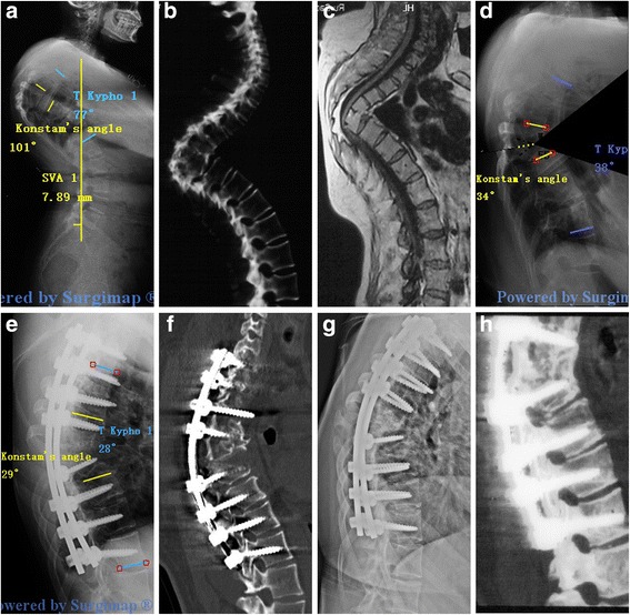
Fig. 5.

A 36-year-old patient with Pott’s deformity. The patient’s main complaints were increasing neurological deficit and cosmetic issues. Pre-operative radiographs (a), CT (b) and MRI (c) show that the apex of kyphosis is located at T6-T9, The Konstam’s angle was 101° and the TK was 77°. The simulation of VCD osteotomy in Surgimap (d). The Konstam’s angle was corrected to 29° and the TK was 28° immediately after the surgery (e, f). 28 months follow-up X-ray (g) and CT (h) scan show a solid fusion
