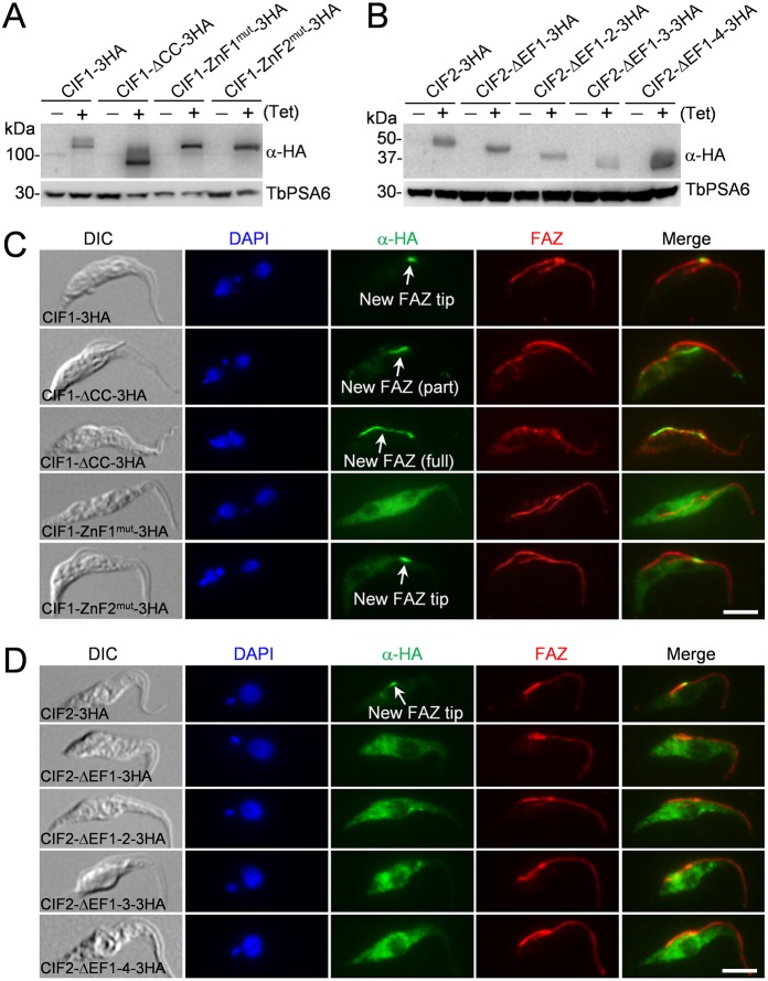
Fig. 4.
Subcellular localization of CIF1 and CIF2 mutants. (A,B) Western blotting to monitor the level of triple HA-tagged wild-type and mutant CIF1 and CIF2, which were induced with 0.1 µg/ml tetracycline for 16 h. TbPSA6 serves as the loading control. (C,D) Immunofluorescence microscopy to examine the subcellular localization of wild-type and mutant CIF1 (C) and CIF2 (D). Cells were co-immunostained with FITC-conjugated anti-HA antibody to label 3HA-tagged CIF1/CIF2 and their mutants and anti-CC2D to label the FAZ filament. DIC, differential interference contrast image. Scale bars: 5 µm.

