Fig. 2.
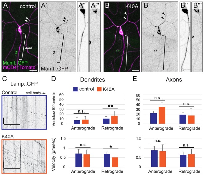
The αTub84B K40A mutation does not affect the polarized distribution of Golgi outposts, but does affect lysosome motility in dendrites. (A–B‴) Golgi outposts, marked by ManII::GFP (green), localize to dendrites in both control (A–A‴) and αTub84BK40A neurons (B–B‴). ManII::GFP (green in A,B; black in A′,A‴,B′,B‴) and CD4::Tomato (magenta in A,B, black in A″,B″) are expressed in class IV da neurons under the control of the ppk enhancer. Bracket, axon; arrowheads, Golgi outposts in dendrites. Scale bars: 25 µm. (C) Representative kymographs of lysosome dynamics in the dendrites of control (top) and αTub84BK40A (bottom) neurons. Scale bar x-axis: 10 µm; scale bar y-axis: 10 s. Lysosomes are marked by Lamp1::GFP. The cell body is to the right. (D,E) In dendrites (D), lysosomes traveling in the retrograde direction in αTub84BK40A neurons display increased flux (top) and reduced velocity (bottom). Lysosome motility in axons (E) is unaffected by the αTub84B K40A mutation. Dendrites (D, flux): 30 wild-type control dendrite segments and 29 αTub84BK40A dendrite segments were analyzed (mean±s.d.); P=0.008. Dendrites (D, velocity): 32 wild-type control dendrite segments and 29 αTub84BK40A dendrite segments were analyzed (mean±s.d.); P=0.012. Axons (E, flux): 7 wild-type control axons and 5 αTub84BK40A axons were analyzed (mean±s.d.). Axons (E, velocity): 7 wild-type control axons and 5 αTub84BK40A axons were analyzed (mean±s.d.). *P=0.01–0.05; **P=0.001–0.01; n.s., not significant (two-tailed Student's t-test).

