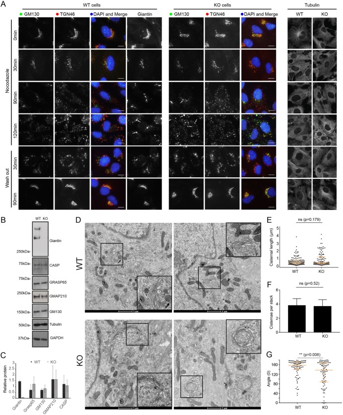
Fig. 3.
Loss of giantin leads to mild changes in Golgi mini-stack structure. (A) Representative maximum projection images of WT and giantin-KO cells incubated with 5 µm nocodazole as indicated and immunolabelled for cis-Golgi (GM130) and trans-Golgi (TGN46) markers or tubulin. In wash-out panels, cells were incubated with nocodazole for 3 h then washed and incubated in growth medium for time indicated. Scale bars: 10 μm. (B) Western blot analysis of golgin expression in WT and KO cells. (C) Quantification of blots represented in B (n=3, mean and s.d. shown). (D) Transmission electron micrographs of WT and KO cells incubated with 5 μM nocodazole for 90 min. Inserts show zoom of region denoted by black squares. (E–G) Quantification of experiments represented in D showing (E) cisternal length, (F) number of cisternae per stack and (G) the angle between lines drawn from each lateral rim of the stack to the centre (n=3; 27 WT and 21 KO cells quantified; E and G show median and interquartile range; F, mean and s.d.; statistics Mann–Whitney).

