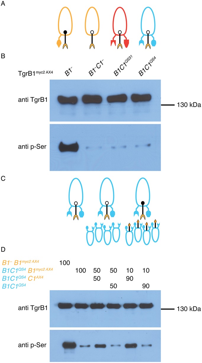
Fig. 5.

Phosphorylation of TgrB1 depends on association with a matching TgrC1. (A) Ovoids represent cells, protrusions represent TgrB1 and TgrC1 proteins, and colors represent allotypes: AX4 (tan), tgrB1C1QS31 (red), tgrB1C1QS4 (blue). The black protrusions represent the extra TgrB1myc2AX4 protein. A solid dot on TgrB1 represents phosphorylation; open dot indicates no phosphorylation. (B) Western blot analysis of proteins pulled down with anti-Myc antibodies from tgrB1-null (B1–), tgrB1-null, tgrC1-null (B1–C1–), tgrB1C1QS31 (B1C1QS31) and tgrB1C1QS4 (B1C1QS4) cells. (C,D) tgrB1C1QS4tgrB1myc2AX4 cells in a pure population and in mixes with tgrB1C1QS4 or with tgrB1C1QS4tgrC1AX4 (C) were used for western blot analysis (D). The mixing ratios (%) are indicated above the lanes. We included the tgrB1–tgrB1myc2AX4 strain as a positive control (left lane).
