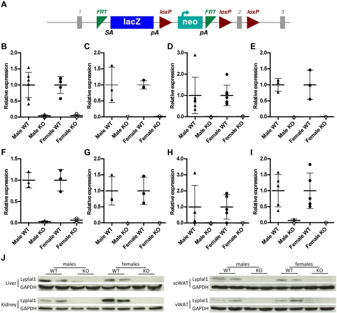
Fig. 1.
Mice were generated with the Lyplal1tm1a allele, resulting in knockout at both the protein and the RNA level. (A) Diagram showing the Lyplal1tm1a allele design (figure obtained from IMPC, www.mousephenotype.org/data/genes/MGI:2385115). RNA and protein were extracted from organs from 28-week-old mice. (B-I) qPCR analysis of Lyplal1 mRNA levels in gastrocnemius (B), heart (C), liver (D), kidney (E), spleen (F), BAT (G), scWAT (H) and vWAT (I). Data are presented as means±s.d. Black triangles, male Lyplal1+/+; white triangles, male Lyplal1tm1a/tm1a; black circles, female Lyplal1+/+; white circles, female Lyplal1tm1a/tm1a. (J) Protein levels of Lyplal1 and GAPDH were determined by Western blot in liver, kidney, scWAT and vWAT lysates. Representative blots are shown. [(B) n=5 female Lyplal1tm1a/tm1a, n=7 other groups; (C,E-G) n=3 each group; (D) n=7 Lyplal1+/+, n=8 Lyplal1tm1a/tm1a; (H) n=8 male Lyplal1+/+, n=6 male Lyplal1tm1a/tm1a, n=5 female Lyplal1+/+, n=5 female Lyplal1tm1a/tm1a; (I) n=5 male Lyplal1+/+, n=3 male Lyplal1tm1a/tm1a, n=5 female Lyplal1+/+, n=6 female Lyplal1tm1a/tm1a; (J) n=3 samples from each sex and genotype per tissue]. KO, knockout; scWAT, subcutaneous white adipose tissue; vWAT, visceral white adipose tissue; WT, wild type.

