Fig. 3.

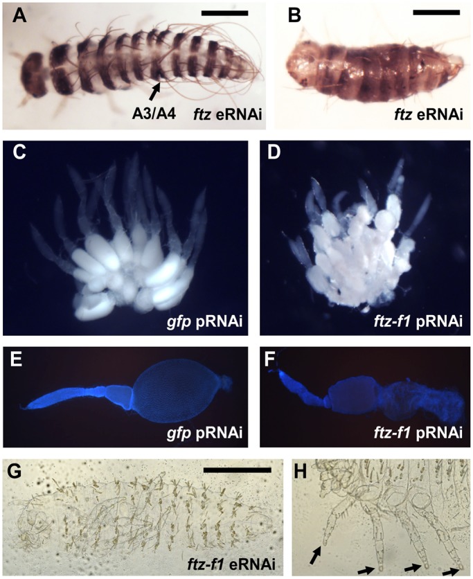
Dmac-ftz and ftz-f1 RNAi. (A,B) Hatched offspring after Dmac-ftz eRNAi. (A) Mildly affected embryo with single segmental fusion (arrow). (B) Severely affected larva with shortened body length and fewer segments. (C,E) Wild-type-like ovaries from gfp dsRNA-injected control female. (C) Large, oval-shaped, developing oocyte in each ovariole. (E) DAPI staining of dissected ovariole reveals large oocyte. (D,F) Ovaries from Dmac-ftz-f1 dsRNA-injected females. (D) Small primary oocytes clustered in each ovariole. No mature oocyte is visible. (F) Dissected ovariole with shrunken oocyte. (G) Unhatched larva from Dmac-ftz-f1 dsRNA-injected female has normal number of segments without any obvious segmentation defect. (H) Arrows indicate truncated distal ends of legs. Scale bars: 500 µm.
