Fig. 2.
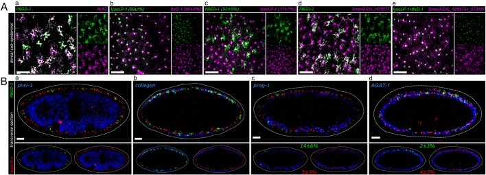
Co-expression analysis of pigment cell markers. (A) z-projections of coronal planes across 20 µm in the dorsal subepidermal layer by FISH. Co-expression is represented as follows: a percentage following a gene name indicates the percentage of cells labeled with this gene which also express the other gene in the same panel. (a) The dendritic markers PBGD-1 and ALAS show a high level of co-expression. (b) The punctate markers lysoLP-1 and thrD-1 show a high level of co-expression. (c) Dendritic+ cells (PBGD-1) highly co-express punctate markers, while a smaller subset of punctate+ cells (lysoLP-1) co-express dendritic markers. (d) Punctate marker-labeled cells possess dendritic cellular morphology. SmedASXL_005875 in situ signals can be detected in the processes of its marked cells. (e) Punctate markers with strict punctate cellular expression patterns (lysoLP-1 and thrD-1) are expressed in the same population that is labeled by punctate markers with process expression (SmedASXL_005875 and SmedASXL_073507). (B) Transverse sections of FISH indicate the localization of pigment cells (marked by PBGD-1 and lysoLP-1) in the subepidermal layer. (a) Pigment cells are located more distal-lateral to the stem cell compartment marked by piwi-1. (b) Pigment cells are interspersed with muscle cells labeled by collagen (SmedASXL_012760). (c) Pigment cells are more distal-lateral to early epidermal progenitor cells marked by prog-1. A small population of dendritic+ cells (14±6%) and punctate+ cells (9±3%) also expresses prog-1. (d) Pigment cells are in the same layer as late epidermal progenitor cells marked by AGAT-1. Scale bars: 50 μm.

