Fig. 2.

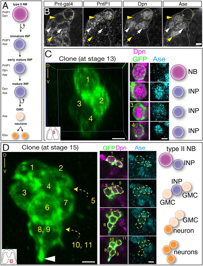
Clonal analysis shows that type II neuroblasts make INPs, GMCs and neurons during embryogenesis. (A) Molecular markers used to identify cell types within type II lineages. (B) Embryonic type II neuroblasts generate embryonic-born INPs and GMCs. Dorsomedial view of a type II neuroblast cluster in a stage 16 embryo showing a type II neuroblast (Pnt-gal4+ PntP1+ Dpn+ and Ase−; yellow circle); an immature INP (Pnt-gal4+ PntP1+ Dpn− and Ase+; yellow arrowhead); mature INP (Pnt-gal4+ PntP1+ Dpn+ and Ase+; white arrowhead); a mature INP that has lost PntP1 expression (Pnt-gal4+ PntP1− Dpn+ and Ase+; white arrow); and a GMC (Pnt-gal4+ PntP1− Dpn− and Ase+; yellow arrow). (C) Single neuroblast clone assayed at stage 13 (location shown in inset): four-cell clone containing a type II neuroblast and three INPs. Orientation is dorsal up, with the neuroblast closest to the dorsal surface of the brain. (D) Single neuroblast clone assayed at stage 15 (location shown in inset): eleven-cell clone containing a type II neuroblast, two INPs, four GMCs and four neurons. Orientation is dorsal up, showing that the neurons are sending projections ventrally (arrowhead). Scale bars: 5 μm (B); 10 μm (C,D, clone projection); 5 μm (C,D, insets). n=1 for each clone shown; n>20 for total clone number analyzed. NB, neuroblast.
