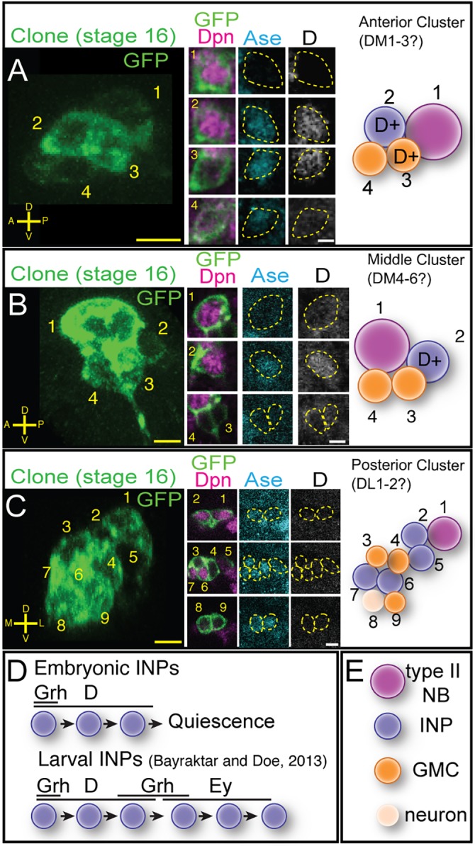
Fig. 6.

Embryonic INPs express the temporal transcription factor Dichaete. (A) Anterior cluster clone containing Dichaete (D)+ INPs. Four-cell FLP-out clone at stage 16 (left) stained for the clone marker (GFP, green), Dpn (magenta), Ase (cyan) and D (white). The clone contains a type II neuroblast (1), a D+ INP (2) and two GMCs, one D+ and one D− (3,4). (B) Middle cluster clone containing D+ INPs. Four-cell FLP-out clone at stage 16 stained as in A containing a type II neuroblast (1), one D+ INP (4) and two D− GMCs (2,3). (C) Posterior cluster clone lacking D+ INPs. Nine-cell FLP-out clone at stage 16 (left) stained as in A containing a type II neuroblast (1), four D− INPs (2,5-7), three D− GMCs (3,4,9) and one D− neuron (8). Scale bars: 7 μm (clonal projections); 5 μm (insets). (D) Model for INP temporal factor expression; top, embryonic INPs from anterior and middle clusters; bottom, larval INP temporal factor expression (Bayraktar and Doe, 2013). (E) Cell type key for panels above. n>20 for stage 16 clones analyzed.
