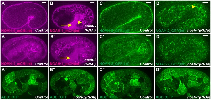
Fig. 3.
NOAH-1 and NOAH-2 require each other for apical secretion. (A-B″) Fluorescent images of control embryos carrying both NOAH-1_mCH(int) and ABD::GFP (A-A″), or the same strain treated with noah-2(RNAi) (B-B″). (C-D″) Fluorescent images of control embryos carrying NOAH-2_GFP(int) (C,C′) or ABD::GFP (C″) markers, and the same strains treated with noah-1(RNAi) (D-D″). The ABD::GFP expression showed some cell-to-cell variation. (A-D) Focal plane through the middle of the embryo; (A′-D″) z-projection. Note the perinuclear accumulation (arrowheads in B,D) of NOAH-1_mCH(int) (B) and NOAH-2_GFP(int) (D), and the extra-embryonic presence of NOAH-1_mCH(int) (arrows in B,B′); the actin pattern was not affected (A″-D″). Scale bars: 5 μm.

