Fig. 6.
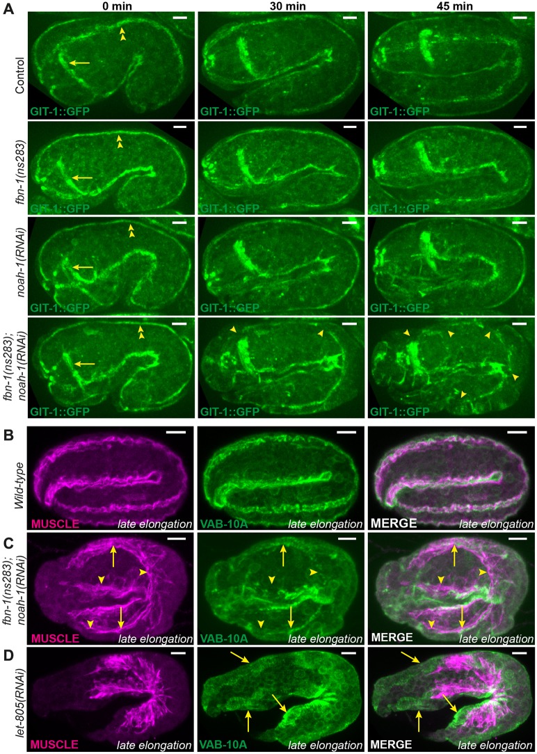
Embryonic sheath protein depletion induces hemidesmosome disruption and muscle detachment. (A) GIT-1::GFP fluorescence time-lapse sequences showing discontinuous CeHDs in an fbn-1(ns283); noah-1(RNAi) embryo beyond the 2-fold stage (n=16/16), which was not observed in fbn-1(ns283) (n=14/14), noah-1(RNAi) (n=6/6) or control (n=6/6) embryos. Arrows indicate nerve ring; double arrowheads, CeHDs; single arrowheads, regions of disrupted CeHDs. (B-D) Wild-type (B), fbn-1(ns283); noah-1(RNAi) (C) and let-805(RNAi) (D) embryos at late elongation stages stained with antibodies against a muscle-specific antigen and the CeHD component VAB-10A, and their merged images. Arrowheads in C indicate regions of disrupted CeHDs. Arrows in C,D indicate regions of CeHDs overlapping with muscle in fbn-1(ns283); noah-1(RNAi) but not in let-805(RNAi) embryos; n≥16. All panels are z-projections. Scale bars: 5 μm.

