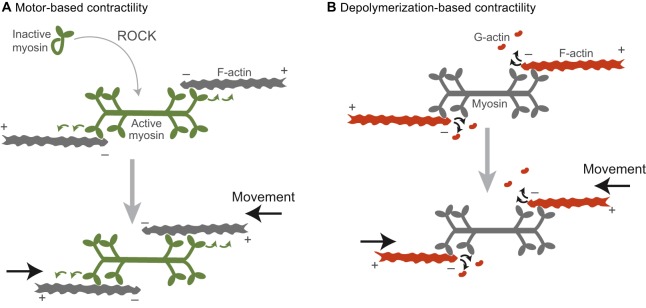
Fig. 1.
Two models by which networks of actin and myosin can generate contractile forces. (A) Myosin is activated by ROCK and polymerizes into a bipolar filament (green). Contractility (black arrows) is generated by the motor activity of myosin as it walks along antiparallel actin filaments (green arrows). (B) Contractility (black arrows) is driven by F-actin depolymerization into G-actin. In this case, myosin would act as one of potentially many crosslinkers between neighboring filaments. The plus (barbed) and minus (pointed) ends of the F-actin filaments are denoted. Note that here, for illustrative purposes, we denote actin subunits depolymerizing from the filament end, but in reality proteins that mediate depolymerization (i.e. cofilin) cause filament severing (Andrianantoandro and Pollard, 2006). Colors indicate the force generator in each model.

