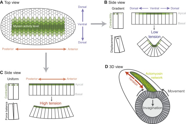
Fig. 4.
A spatial pattern of contractility results in Drosophila ventral furrow formation. (A) Ventral surface of the embryo. The distribution and magnitude of myosin activity is represented in green (darker green represents more myosin activity). (B) An apical contractility gradient causes a force imbalance that allows cells to constrict and the tissue to bend along the dorsal-ventral axis. (C) Uniform apical contractility along the anterior-posterior axis promotes balanced forces and tension, which prevents tissue bending along this axis. (D) An embryo in the process of folding, with the orientation of actomyosin fibers illustrated in yellow.

