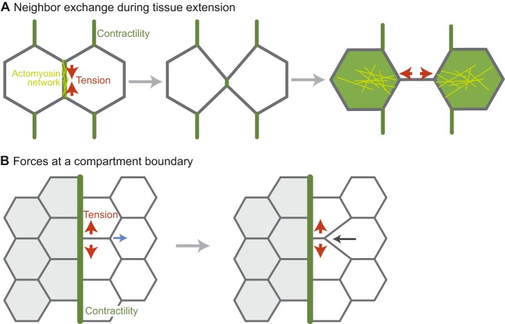
Fig. 6.
Polarized junctional actomyosin contractility during tissue extension and at compartment boundaries. (A) Neighbor exchange during tissue extension. Actomyosin is planar polarized to the vertical junction. This actomyosin network leads to apical junction shrinkage either through contraction or by directionally stabilizing fluctuations in junction length (Rauzi et al., 2010). The junction is then expanded in the horizontal direction by medial actomyosin contractility (in the green cells). (B) The forces at a compartment boundary that inhibit cell rearrangement. When a rearrangement occurs near the boundary, the boundary resists deformation. For example, the horizontal junction shrinks from one end so that the boundary remains straight. Regions of contractility are depicted in green, with network organization in yellow. Red arrows denote the direction of contractile tension. The blue arrow denotes the direction of low tension and the black arrow denotes the direction of movement as a result of the low tension.

