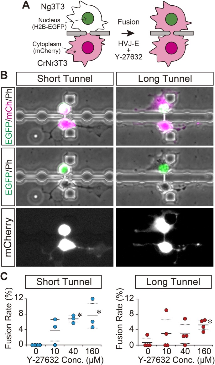
Fig. 2.
Promotion of strictured cytoplasmic connection between live single cells. (A) Schematic illustration of the experimental procedure. Cell fusion was induced through a microtunnel between heterogeneous pairs of Ng3T3 and CrNr3T3 cells by exposure to fusion medium with 160 μM Y-27632 for 1 h. Cell fusion was detected by transfer of mCherry from CrNr3T3 to Ng3T3 cells. (B) Achieved strictured cytoplasmic connection. Data pertaining to short and long tunnels are presented. A strictured cytoplasmic connection with a length corresponding to that of the microtunnel was successfully obtained. H2B-EGFP (EGFP: nuclei), mCherry (mCh: cytoplasm) and phase contrast (Ph) images are shown in green, magenta (or white) and gray, respectively. (C) Effect of Y-27632 supplementation on cell fusion through a microtunnel. Fusion rate in heterogeneous cell pairs of Ng3T3 and CrNr3T3 cells are presented. Each plot represents the result from one independent experiment. Horizontal solid and dashed lines represent average values and ±s.d., respectively. *Significantly different from 0 μM evaluated by Dunnett's test (P<0.05).

