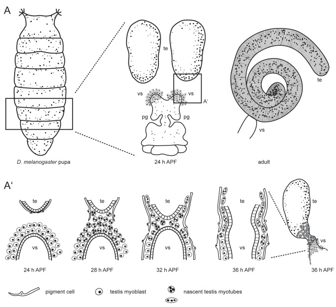
Fig. 1.
Scheme of Drosophila testis myotube migration. (A) The male reproductive tract develops during metamorphosis. At 24 h APF, the single genital disc and paired testes (te) are separate organs. The seminal vesicles (vs) and the paragonia (pg) already start to grow. In the adult, the tubular testis is connected to the seminal vesicle. (A’) During metamorphosis, the prospective seminal vesicles and testes grow towards each other and fuse. On genital discs 24 h APF, testis-relevant myoblasts accumulate on the prospective seminal vesicle. Pigment cells cover the pupal testis. At 28 h APF, myoblasts fuse to build multinucleated testis myotubes. These nascent testis myotubes migrate beneath the pigment cells onto the pupal testis, while pigment cells migrate from the testis onto the developing seminal vesicle. By 36 h APF, the epithelia of seminal vesicles and the terminal epithelium of the testes have fused. Modified after Bodenstein (1950), Kozopas et al. (1998), Kuckwa et al. (2016).

