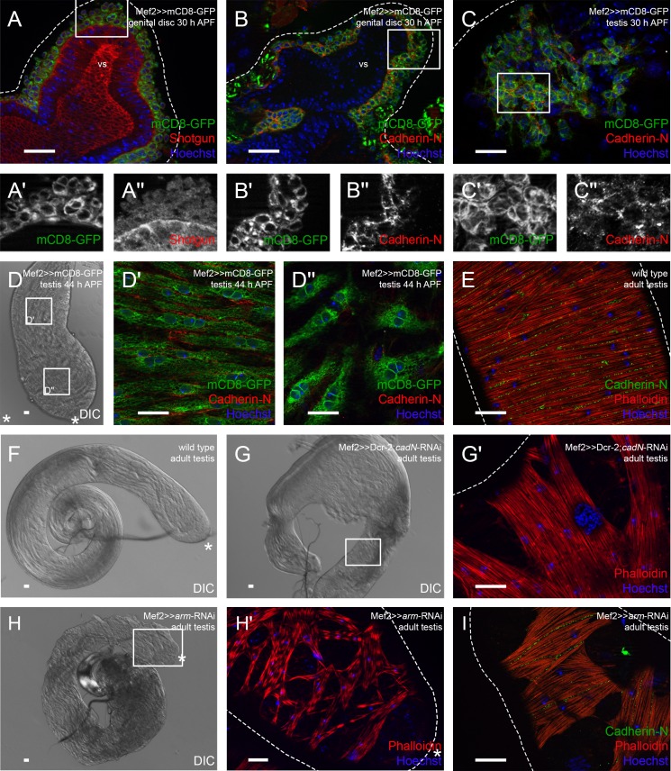
Fig. 2.
Knock-down of Cadherin-N or Armadillo strongly reduces the adhesion between testis myotubes. Immunofluorescence analyses of genital discs and testes. (A) Seminal vesicles 30 h APF stained or marked with anti-Shotgun (red), GFP (green; myoblasts and myotubes on genital discs and pupal testes marked with Mef2≫mCD8-GFP), and Hoechst (blue; nuclei). (A′,A″) Enlargement of boxed area in A, stained or marked as indicated. (B) Genital discs 30 h APF and (C) testis 30 h APF stained or marked with anti-Cad-N (red), GFP (green), and Hoechst (blue), magnification of prospective seminal vesicle is shown. (B′,B″,C′,C″) Enlargement of boxed area in B and C stained or marked as indicated. (D–D″) Testis 44 h APF. (D) Differential interference contrast (DIC) micrograph of testis 44 h APF, (D′,D″) enlargement of boxed area in D stained or marked with anti-Cad-N (red), GFP (green), and Hoechst (blue). (E) Adult testis stained with Hoechst (blue), Phalloidin to visualize F-actin (red), and anti-Cad-N (green). (F) DIC micrograph of wild-type testis. (G) DIC micrograph of cad-N knock-down testis; (G′) enlargement of boxed area in G showing Phalloidin (red) and Hoechst (blue) staining of testis muscle sheath. (H) DIC micrograph of arm knock-down testis; (H′) enlargement of boxed area in H showing Phalloidin (red) and Hoechst (blue) staining of testis muscle sheath. (I) Adult arm knock-down testis stained with Hoechst (blue), Phalloidin to visualize F-actin (red), and anti-Cad-N (green). Dotted lines reflect approximate shape of the organ. Asterisk, hub region; vs, seminal vesicle. Scale bars: 20 µm.

