Fig. 6.
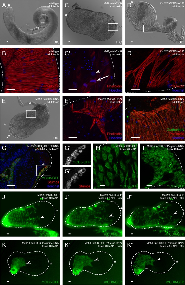
Ths-activated Heartless is essential for populating the testis with myotubes. Analysis of htl and stumps knock-down and ths mutant. (A) DIC micrograph of adult wild-type testis. (B) Phalloidin staining to visualize F-actin (red), and Hoechst staining of nuclei (blue). (C) DIC micrograph of adult htl knock-down (v6692) testis. (C′) Enlargement of boxed area in C showing adult htl knock-down testis stained with Phalloidin (red; F-actin) and Hoechst (blue; nuclei); arrowhead, pigment cell nuclei; arrow, spermatids during individualization. (D) DIC micrograph of adult ths mutant testis. (D′) Enlargement of boxed area in D showing adult ths mutant testis stained with Phalloidin (red) and Hoechst (blue). (E) DIC micrograph of adult stumps knock-down testis. (E′) Enlargement of boxed area in E showing adult stumps knock-down testis stained with Phalloidin (red) and Hoechst (blue). (F) Adult stumps knock-down testis stained with anti-Cad-N (green), Phalloidin (red), and Hoechst (blue). (G-G″) htl knock-down genital disc 24 h APF stained or marked with anti-Stumps (red), GFP (green), and Hoechst (blue); magnification of prospective seminal vesicle is shown. (G′,G″) Enlargement of boxed area in G marked with GFP or stained with anti-Stumps. (H) Myoblasts on wild-type testis 40 h APF marked with Mef2-driven mCD8-GFP (green). (I) Myotubes of stumps knock-down testis 40 h APF marked with GFP (green). (J-K″) Live imaging over time of testes 40 h APF expressing Mef2-driven mCD8-GFP to reveal the migration of nascent myotubes in an ex vivo culture of (J-J″) wild-type testis and (K-K″) stumps knock-down testis. Dotted lines reflect the approximate shape of the organ. Arrowheads, the front of migrating nascent myotubes; asterisk, hub region; vs, seminal vesicle. Scale bars: 20 µm.

