Fig. 3.
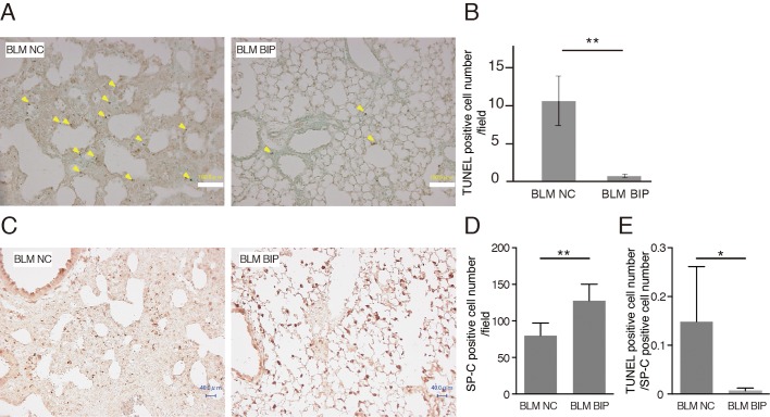
BIP-V5 decreases apoptosis during bleomycin-induced pneumopathy. (A) Representative images after TUNEL staining of the lung tissues from the BLM+NC and BLM+BIP groups on day 7. The arrowheads indicate evidence of apoptosis, including chromatin condensation. Scale bars: 100 µm. (B) Quantitative results of the number of TUNEL-positive cells in the lung tissues. (C,D) Immunohistochemical analysis of SP-C expression (C) and the quantitative results of SP-C-positive cell number (D) on day 7. TUNEL-positive cells and SP-C-positive cells were counted in all fields (40–55 fields) of the lungs using a light microscope with 200× magnification. Scale bars: 40 µm. (E) The ratio of TUNEL-positive cells to SP-C-positive cells. Data are presented as the mean ±s.e.m. from five mice per group. *P<0.05, **P<0.01.

