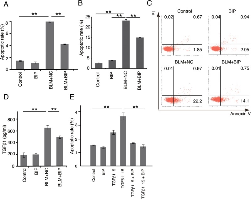
Fig. 5.
Effect of BIP-V5 on the apoptosis of pulmonary alveolar epithelial cells. (A) The apoptotic rate of A549 cells under BLM treatment. A549 cells were treated with normal saline or BLM (120 mU/ml) for 24 h. BIP-V5 (200 µM) was pre-dosed 1 h before the administration of saline or BLM. (B,C) The apoptotic rate of LA-4 cells under BLM treatment. LA-4 cells were treated with normal saline or BLM (120 mU/ml) for 12 h. BIP-V5 (200 μM) was pre-dosed 1 h before the administration of saline or BLM. Apoptotic rate (B) was measured by flow cytometry (C) as Annexin V positivity. (D) A549 cells were treated for 24 h with BLM. BIP-V5 (200 µM) was pre-dosed 1 h before the administration of BLM. TGF-β1 concentration in the supernatant of the medium was measured by ELISA. (E) The percentage of apoptotic cells under TGF-β1 stimulation. A549 cells were treated with normal saline or TGF-β1 (5 ng/ml or 15 ng/ml) for 72 h. BIP-V5 was pre-dosed 1 h before the administration of saline or TGF-β1. Data were collected from three separate experiments and are expressed as the mean±s.e.m. **P<0.01. PI, propidium iodide.

