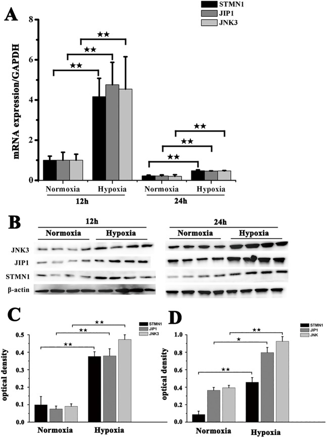
Fig. 3.
Expression of STMN1, JIP1 and JNK3 in muscle fibroblasts from NMRs before and after exposure to hypoxia. (A) Real-time PCR analysis of STMN1, JIP1 and JNK3 mRNA expression in muscle fibroblasts before and after exposure to hypoxia (5% O2) for 12 h or 24 h. (B) Western blot detection of STMN1, JIP1 and JNK3 protein expression in fibroblasts before and after exposure to hypoxia (5% O2) for 12 h or 24 h. β-actin was used as an internal loading control. (C,D) Band density analysis of STMN1, JIP1 and JNK3 protein expression in fibroblasts before and after exposure to hypoxia (5% O2) for (C) 12 h or (D) 24 h. Data represent the mean±s.e.m. of five independent experiments. ★P<0.05, ★★P<0.01 (t-test).

