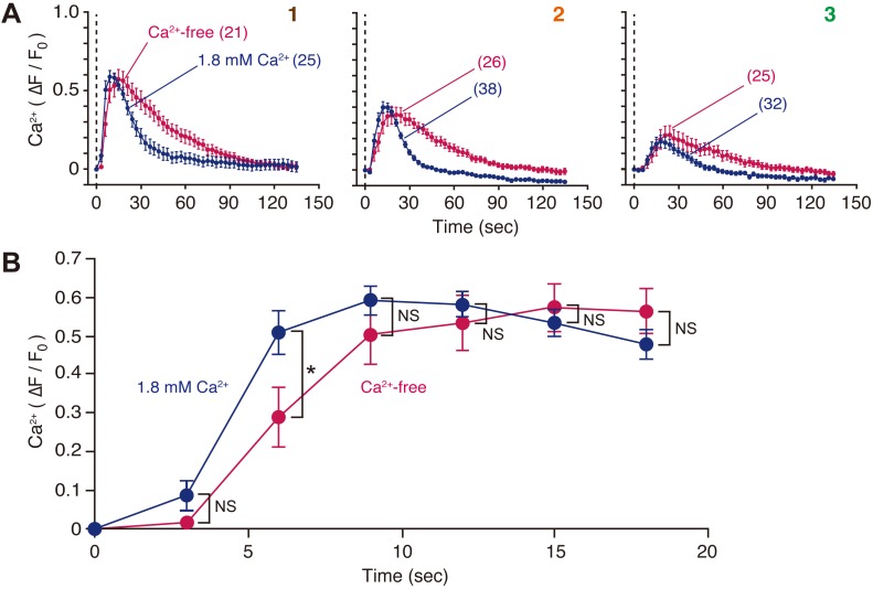
Fig. 4.
Cell membrane disruption induces Ca2+ mobilization in neighboring MDCK cells. (A) Cells loaded with Calcium Green-1 AM were wounded at time zero with a glass needle in the presence or absence of extracellular Ca2+, and changes in fluorescence intensity in the cytoplasmic region were compared. Cells were numbered as per Fig. 2A. The number of observed cells is indicated in parentheses. See also Movie 2. (B) To compare the initial phase of increase in [Ca2+]i, data from cell #1 in A were expanded. *P=0.0333; NS, not significant.

