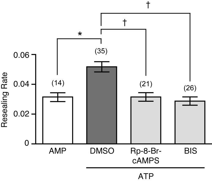
Fig. 6.
Potentiation of membrane resealing induced by ATP is suppressed by PKA and PKC inhibitors in MDCK cells. Cells were pretreated for 10 min with either kinase inhibitors or DMSO. Then, cells were wounded with a glass needle after the addition of 100 µM ATP in the presence or absence of inhibitors, respectively, and the resulting resealing rates were analyzed. As a control, cells treated with 100 µM AMP were wounded with a glass needle. Resealing rates were analyzed 5–20 min after the addition of nucleotides. The number of observed cells is indicated in parentheses. *P=0.0007; †P<0.0001.

