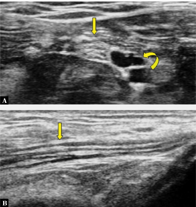
Fig. 2.
A transverse view of the tibial nerve, showing a honeycomb pattern (straight arrow in A) due to hypoechoic areas (nerve fascicle groups) distributed over a hyperechoic background (perineurium). The echopoor areas are posterior tibial vessels (curved arrow in A). In the longitudinal view, the nerve appears as the long, slim structure with alternate hypoechoic and hyperechoic stripes (Straight arrow in B).

