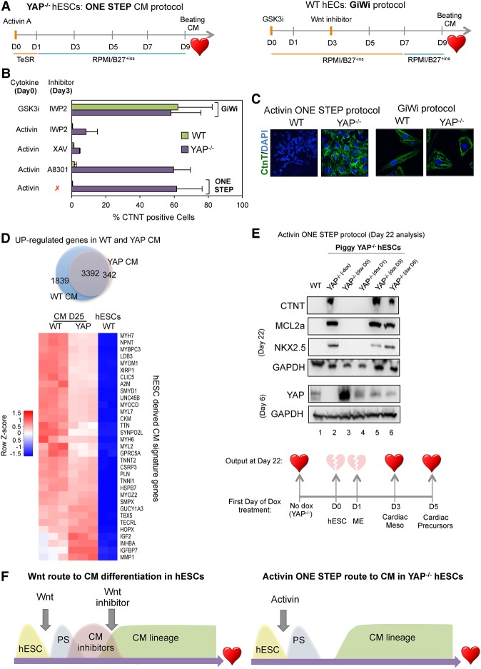
Figure 7.
Human cardiomyocyte differentiation using a one-step protocol. (A) The schematic outline of the one-step protocol used to differentiate YAP knockout hESCs into beating cardiomyocytes in comparison with the GiWi protocol. (B) Wild-type and YAP knockout cells were differentiated using the conditions indicated at the left of the graph. Around day 22 following the initial treatment, cells were stained with the cardiac marker CTNT and subjected to FACS analysis. Mean (SD). n ≥ 3. (C) Immunostaining of wild-type- and YAP knockout-derived cardiomyocytes using the protocol indicated below the images. (D) The Venn diagram shows up-regulated genes in wild-type and YAP knockout cardiomyocytes compared with hESCs. (Below) The heat map shows a list of up-regulated cardiac markers in wild-type and YAP−/− cardiomyocytes. (E) Immunoblot analysis of cardiac markers CTNT, NKX2.5, and MCL2a in wild-type and PiggyYAP cells at day 22 after initial differentiation according to the Activin one-step protocol. Doxycycline was added at different time points (not added, day 0, day 1, day 3, or day 5). Note that the reintroduction of YAP at day 0 and day 1 impaired Activin-mediated differentiation, whereas expression of YAP in later stages (day 3 and day 5) did not affect differentiation, as indicated in the bottom illustration. (F) A schematic diagram summarizing the results of the figure. (CM) Cardiomyocyte.

