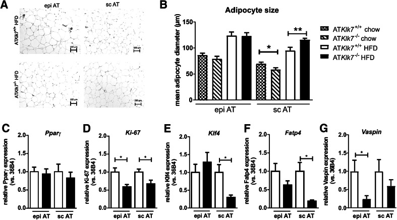
Fig. 5.
Adipose tissue morphology and adipocyte size were altered in ATKlk7 −/− mice. a Histological analyses by H&E staining of epigonadal AT (left panel) and subcutaneous AT (right panel) suggest hypertrophy of subcutaneous adipocytes of ATKlk7 −/− compared to control mice after HFD. b Mean epigonadal (epi) and subcutaneous (sc) adipocyte diameters were analyzed using a multisizer (Beckman Coulter) and confirmed HFD-induced hypertrophy of subcutaneous adipocytes. The mean size of epigonadal adipocytes was not different between the genotypes, both under chow and HFD conditions, whereas expandability of sc AT seems to be improved in ATKlk7 −/− compared to control mice (n = 9–14). c–g Expression of Pparγ, Ki-67, Klf4, Fatp4 and vaspin mRNA was examined by quantitative RT-PCR (n = 7–8). Data are represented as mean ± SEM and for each AT depot differences between genotypes were tested for statistical significance by a two-tailed Student’s t test; *P < 0.05, **P < 0.01

