
Figure 2. Membrane Structure and Pore Size Distribution.
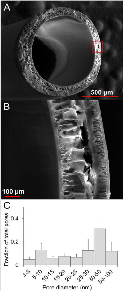
Transverse SEM micrographs (A and B) illustrate the structure of HFM’s, which consist of nano-porous interior and exterior skin layers that encapsulate an asymmetric micro-porous (size range 1–500um) interior structural layer. Evapoporometric characterization of the skin layer pore diameter distribution (mean = 39.8 nm ± 3.9 SD, n = 3) revealed a predominantly mesoporous (size range = 1–100nm) distribution of pores with sizes large enough to permit nutrient transport, but small enough to trap large structural proteins within the HFM lumen.