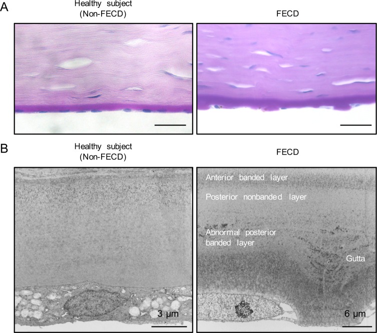
Fig 1. Histology of excrescences of Descemet’s membrane in patients with FECD.
(A) Representative PAS staining images of a healthy donor cornea (left).Representative PAS staining images of a cornea obtained from a patient with FECD. Excrescences, which are clinically called guttae”, were observed on Descemet’s membrane of the patient with FECD. Scale bar: 50 μm (right). (B) Ultra structural analysis of Descemet’s membrane of non-FECD donor cornea was observed using TEM. Scale bar: 3 μm (left). Ultrastructural TEM analysis of Descemet’s membrane of a cornea obtained from a patient with FECD. Flattened CECs adhered to the excrescences on Descemet’s membrane. Scale bar: 6 μm (right).

