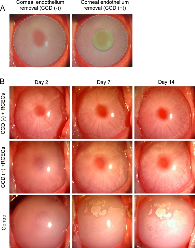
Fig 2. Rabbit corneal endothelium cell (RCEC) injection in the rabbit corneal endothelial dysfunction model with removal of a small central part of Descemet’s membrane.
(A) The corneal endothelium was totally scraped from the Descemet’s membrane with a 20-gauge silicone needle, leaving the remaining Descemet’s membrane intact. These eyes were used as the circular descemetorhexis (CCD) (-) group (left). As the CCD (+) group, a 4-mm diameter of Descemet’s membrane was removed by CCD following total corneal endothelium removal. The gray area indicates the area where the corneal endothelium was removed. The green area indicates the area where Descemet’s membrane was removed. (B) A total of 5.0×10⁵ RCECs, suspended in 200 μl of medium supplemented with 100 μM Y-27632 ROCK inhibitor, was injected into the anterior chamber of the (CCD (-) and CCD (+) groups) (n = 6). Six eyes from which the corneal endothelium was totally removed and Descemet’s membrane remained intact were used as controls. Corneal transparency was restored by intracameral injection of RCECs both in CCD (-) and CCD (+) groups, while control eyes exhibited hazy corneas due to corneal endothelial dysfunction.

