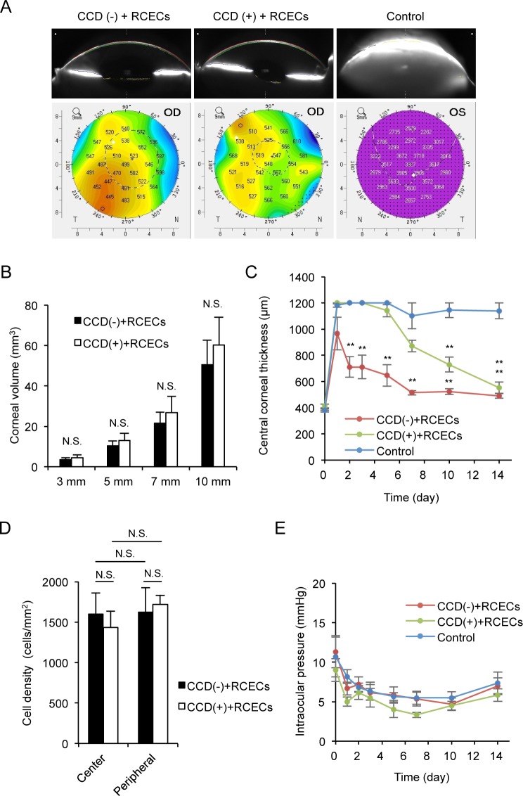
Fig 5. Assessment of the effect of circular descemetorhexis (CCD) on clinical parameters.
(A) Scheimpflug images showed the restoration of an anatomically normal cornea in both the CCD (-) and CCD (+) models at 14 days after surgery. Control eyes showed corneal edema due to corneal endothelial dysfunction. A color map of corneal thickness showed that corneal thickness was thinner in both the CCD (-) and CCD (+) models when compared to the control. (B) The corneal volume was similar, when measured with a PentacamTM instrument at 3, 5, 7, and 10 mm diameters, in both the CCD (-) and CCD (+) models (n = 6). The corneal volume of the control group is not shown, as it was not evaluated with the PentacamTM instrument due to severe corneal edema. N.S. indicates no statistical significance. (C) The central corneal thickness was evaluated with an ultrasound pachymeter for 2 weeks after rabbit corneal endothelium cell (RCEC) injection. The eyes of the CCD (-) and CCD (+) models showed recovery of the central corneal thickness to an almost normal range, whereas this thickness did not recover in the controls. However, recovery of corneal thickness was slower in the CCD (+) model than in the CCD (-) model (n = 6). **P < 0.01, *P < 0.05. (D) Cell density of the regenerated corneal endothelium was determined by analyzing immunofluorescence staining images using ImageJ software. The average cell density of the restored corneal endothelium was similar for the CCD (-) and CCD (+) models in both the central and peripheral areas (n = 6). N.S. indicates no statistical significance. (E) Intraocular pressure (IOP) was evaluated with a Tonovet® instrument, and no abnormal IOP elevation was observed in any of the groups (n = 6).

