
Fig 1.
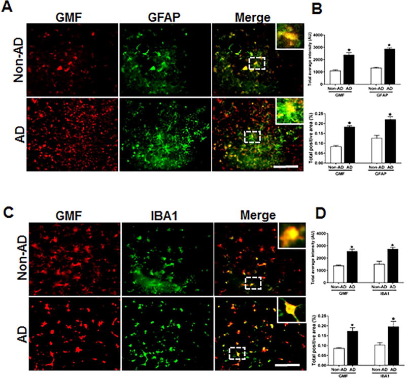
Co-localization of GMF with GFAP and IBA1 in the temporal cortex of AD and non-AD control brain. (A) Sections were immunostained with ant-GMF and anti-GFAP respectively. Representative images showed higher expression and co-localization of GMF (red) with GFAP (green), which clearly indicates astrocytes are one of the site of expression of GMF in human AD brain. (C) Sections were immunostained with anti-GMF and anti-IBA1 respectively. Representative image display the higher expression of and co-localization of GMF(red) with IBA1(red) which clearly indicates microglia are another site in addition to the astrocyte and some neuronal cell for the GMF expression in human AD brain. The values are expressed as mean ± standard error of determination from each group (n= 3–5). *p <0.05 versus non-AD was considered statistically significant. Boxed area shows the enlarged view of co-localization of GMF (red) with GFAP (green) and IBA1 (red) Scale bar = 50μm and AU (Arbitrary Unit). (B&D) Quantification of GMF, GFAP and IBA1based on average labeled intensity and labeled positive area in AD and non-AD brains.