
Fig 4.
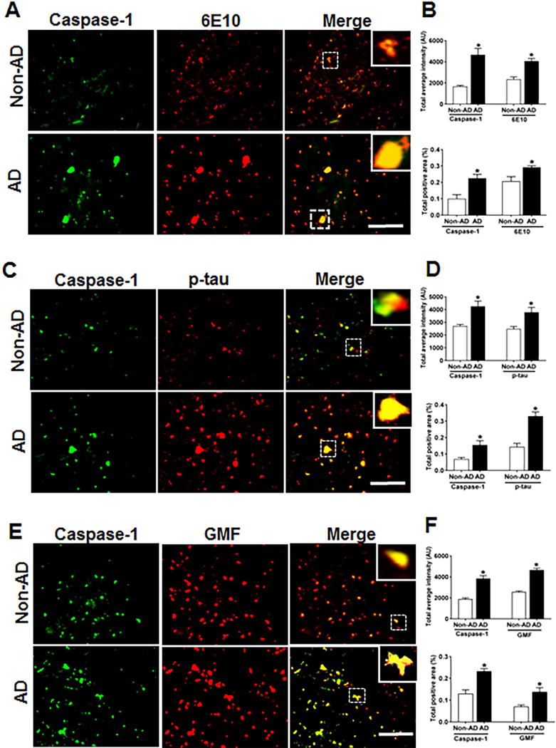
Co-localization of caspase-1 with Aβ, p-tau and GMF in the temporal cortex of AD and non-AD brain. (A) Sections were immunostained with anti-caspase-1 and 6E10 respectively. Representative immunofluorescence staining display the higher expression and co-localization of caspase-1 (green) and 6E10 (red) in AD as compared with non-AD. (C) Sections were immunostained with anti-caspase-1 and anti-p-tau respectively to check the co-localization of caspase-1 with p-tau. Representative immunofluorescence staining displayed the higher expression and co-localization of caspase-1(green) and p-tau (red) in AD compared with non-AD. (E) Sections were immunostained with anti-caspase-1 and anti-GMF respectively to check the co-localization of caspase-1with GMF. Representative immunofluorescence staining displayed the higher expression and co-localization of caspase-1(green) and GMF (red) in AD compared with non-AD. The values are expressed as mean ± standard error, from each group (n= 3–5). *p <0.05 versus non-AD was considered statistically significant. Zoom of boxed area showed the enlarge view of co-localization of caspase-1 (green) with 6E10 (red), p-tau (red) and GMF (red) Scale bar = 50 μm and AU (Arbitrary Unit). (B, D, F) Quantification of Caspase-1, 6E10, p-tau and GMF based on average labeled intensity and labeled positive area in AD and non-AD brains.