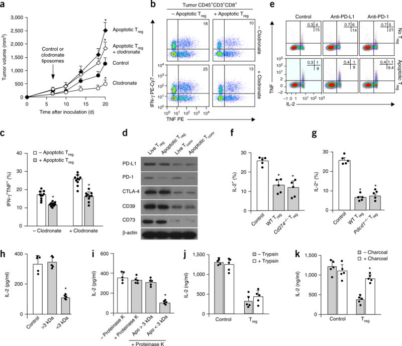
Figure 3.

Apoptotic Treg cells mediate immunosuppression via soluble factor(s). (a–c) The effect of macrophage depletion on apoptotic Treg cell–mediated suppression. Mice bearing MC38 and apoptotic Treg cells were treated with clodronate-loaded liposomes. The plots show tumor volume (a) and numbers of tumor-infiltrating effector T cells (b,c). Data in a and c are the mean and s.d. of n = 10 mice per group. *P < 0.05 versus the control, Student’s t-test. (d) The expression of immunosuppression-associated molecules in live and apoptotic T cell subsets. The indicated mouse T cell subsets were immunoblotted with antibodies to the indicated proteins. (e) The effect of PD-L1 or PD-1 blockade on apoptotic Treg cell–mediated T cell suppression. Apoptotic Treg cells and apoptotic conventional T cells were induced by irradiation. Apoptotic Treg cells and apoptotic conventional T cells were cultured with normal T cells at a ratio of 1:2 in the presence of anti-CD3. We measured the expression of T cell TNF and IL-2 by flow cytometry. (f,g) Immunosuppression mediated by PD-L1−, PD-1−, and wild-type (WT) apoptotic Treg cells. Apoptotic Treg cells from Cd274−/− (f), Pdcd1−/− (g), and wild-type mice were cultured with normal T cells at a ratio of 1:2 in the presence of anti-CD3. We measured the expression of T cell TNF and IL-2 by flow cytometry. Data shown are the mean and s.e.m. (n = 5 mice). *P < 0.05 compared with controls, Wilcoxon test. (h) Immunosuppression mediated by low-molecular-weight components in apoptotic Treg cell–derived supernatants. Treg cells were treated with anti-Fas mAb for 6 h. Treg cell supernatants were passed through a 100-kDa cutoff filter and divided into two fractions with a 3-kDa cutoff filter. The two fractions were collected for T cell immunosuppression assays. T cell IL-2 was detected on day 3 by ELISA. The data shown are the mean and s.d. (n = 5 mice). *P < 0.05 versus the control, ANOVA with Dunett’s post-test. (i,j) Immunosuppression mediated by nonprotein elements in apoptotic Treg cell supernatants. Apoptotic Treg cell supernatants (apo) were treated with proteinase K (i) or trypsin (j) and subsequently used for immunosuppression assays. T cell IL-2 was detected on day 3 by ELISA. The data shown are the mean and s.d. (n = 5 mice). ANOVA with Dunett’s post-test, *P < 0.05. (k) The effect of the removal of small lipid-like factors on immunosuppression mediated by apoptotic Treg cell supernatants. Apoptotic Treg cell supernatants were mixed with charcoal-treated dextran to remove small lipid-like molecules and were subsequently used for immunosuppression assays. T cell IL-2 was detected on day 3 by ELISA. The data shown are the mean and s.d. (n = 5 mice). Wilcoxon test, *P < 0.05 versus the control. Data are representative of 10 (d) or 5 (e) experiments. In b and e, numbers in the upper right show the percentage of cells in the indicated quadrant.
