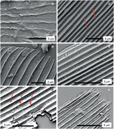
Fig. 4. Detailed SEM images of selected scales shown in Fig. 3.

Labeling same as in Fig. 3. (A) Detail of a type I scale showing herringbone pattern. (B) Detail of a type II scale showing oblique crests at the apical margin. (C to E) Type II scales showing hollow structure, perforations, cross-ridges, and microribs (arrows in Fig. 3, C and D). (F) Detail of a fringed scale of indeterminate origin showing distinct apical margin.
