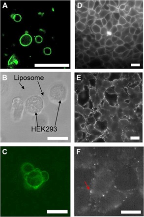
Fig. 3. Delivery of pcNRs to HEK293 cells.

(A) Fluorescence of NR-loaded GUVs. (B and C) Bright-field (B) and fluorescence (C) images of pcNR-loaded GUV fused with the cell membrane. (D) Fluorescence image of HEK293 cells stained with ANEPPS (control). (E and F) pcNRs targeted to membranes at high (E) and low (F) concentrations. Scale bars, 10 μm.
