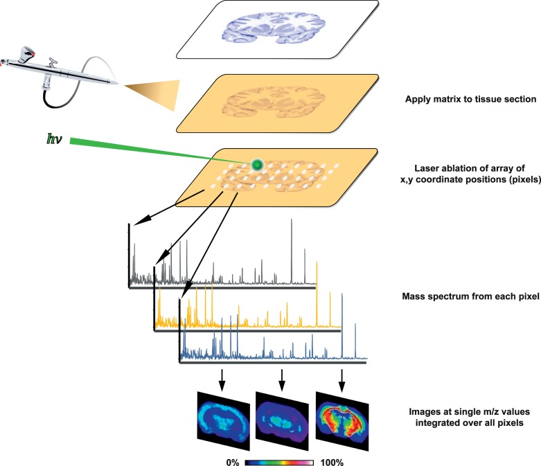
Figure 1.
MALDI IMS process overview. Thin tissue sections are mounted on MALDI target plates and coated with chemical matrix that assists laser desorption and ionization of tissue matrix. As a laser is rastered through a predefined array of coordinate locations (pixels), each location is associated with a unique mass spectrum. Ion signal intensities are encoded as different hues in a color lookup table, and images are created by plotting signal intensities of ions of interest by pixel location. Data-mining approaches are used to discover discriminant spectral features.

