Figure 2.
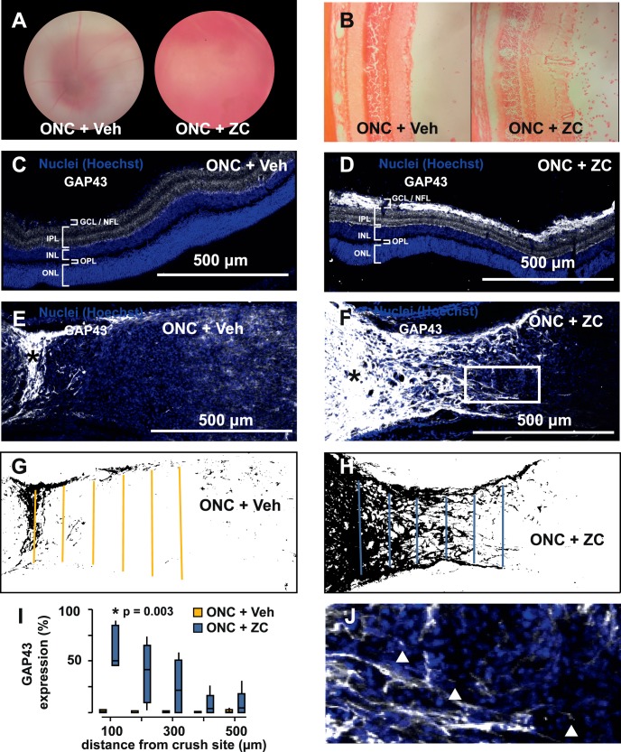
Establishment of an inflammation-induced optic nerve regeneration model. (A) Hazy fundus view 3 days after ONC immediately followed by intravitreal injection of Zymosan A plus CPT-cAMP (ONC+ZC) is caused by an inflammatory infiltrate that is evident on H&E (B). (C, D) GAP43 is elevated in the ganglion cell layer and nerve fiber layer of the ONC+ZC group (day 14). (E, F) The optic nerve distal to the crush site (*) has increased GAP43 expression in the ONC+ZC group (day 14). (G, H) Binarized versions of the images in E and F and linear ROIs drawn at 100-μm intervals were used to quantify GAP43 expression. (I) GAP43 expression as a function of distance from the crush site. (J) Expanded view of the inset box from panel F shows examples of growth cone–like structures that tip GAP43-positive processes in the ONC+ZC group (triangles). ONL, outer nuclear layer; OPL, outer plexiform layer; INL, inner nuclear layer; IPL, inner plexiform layer; GCL/NFL, ganglion cell layer/nerve fiber layer.

