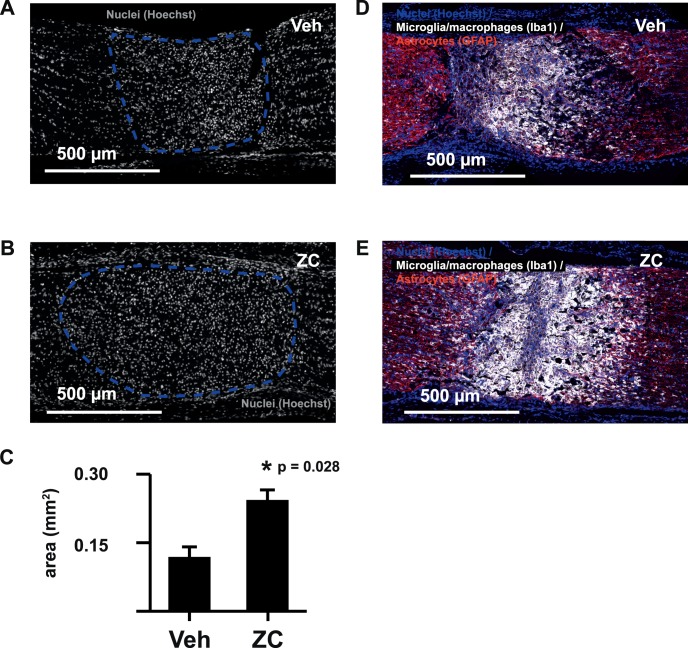
Figure 4.
IIR enhances microglia/macrophage accumulation at the crush site. The size of the hypercellular crush core region (dashed lines in A and B) demonstrated by Hoechst nuclear stain is significantly increased by IIR at day 7 (C). The cellular infiltrate occupying the crush core region strongly labels with microglia/macrophage marker Iba1 (D, E).

