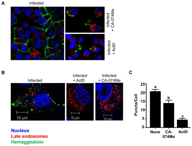
Figure 4. CA-074Me inhibits surface expression of HA protein in A549 cells.
A. A549 cells adhered to coverslips were infected for six hours at an MOI of 10 in the presence of PBS (left), CA-074Me (150 μM; top right), ActD (5 μg/mL; bottom right). About 6 h post-infection, cellular compartments were labelled for late endosomes/lysosome (Lysotracker Red®), HA (green) and nucleus (Hoechst 33342; blue), as described in Methods. Cells were then viewed using a Zeiss LSM 510 confocal fluorescence microscope and images shown are representative cells from respective treatments. B. Similarly, cells were processed as in A, except that cells were infected with IAV-PR8 at MOI of 5 and permeabilized with 0.25% Triton X-100 before HA labelling. Images shown are representative images from three independent experiments. C. The numbers of puncta per cell in random fields of view were quantified and plotted for each treatment group. Data are expressed as means ± SEM (p > 0.05; one-way ANOVA; n=3). Columns accompanied by the same letter are not significantly different from each other by Tukey’s post hoc test.

